-
Follow Us On:
-
-

Intravascular Imaging
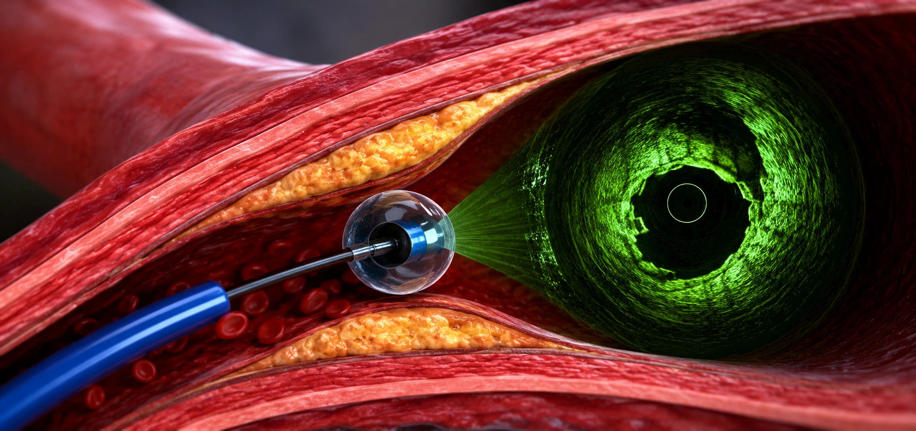
Intravascular Imaging (IVUS and OCT) in Basaveshwaranagar, Bangalore
Intravascular imaging uses advanced catheter-based technology to look inside the coronary arteries with far greater detail than a standard angiogram. It helps cardiologists accurately assess blockages, plan the right treatment, and confirm precise stent placement, especially in complex or high-risk cases.
Key Intravascular Imaging Techniques We Offer
-
IVUS (Intravascular Ultrasound): Uses a tiny ultrasound probe inside the artery to create cross-sectional images that reveal plaque size, composition, and vessel dimensions.
-
OCT (Optical Coherence Tomography): Uses light-based imaging to produce extremely high-resolution pictures of the artery wall, ideal for evaluating plaque detail and stent positioning.
Why Intravascular Imaging Matters
-
Provides a detailed view beyond what a standard angiogram shows.
-
Helps select the correct stent size and length.
-
Confirms complete and accurate stent placement.
-
Reduces the risk of future stent-related complications.
-
Improves outcomes in complex coronary interventions.
Who May Benefit From Intravascular Imaging
-
Patients with complex or heavily calcified blockages.
-
Patients undergoing left main or bifurcation PTCA.
-
Patients with a history of stent-related problems.
-
Patients where blockage severity is unclear on angiogram.
-
Patients requiring precise, image-guided angioplasty.
Benefits of Image-Guided Coronary Treatment
-
Greater accuracy in diagnosing artery blockages.
-
Improved long-term success of stenting procedures.
-
Lower risk of stent under-expansion or malapposition.
-
Better treatment planning for complex cases.
-
Enhanced safety during high-risk coronary procedures.
Book an Intravascular Imaging Consultation
Speak with our interventional cardiology team to understand how IVUS or OCT guided imaging can improve the accuracy and safety of your coronary procedure. Precise imaging leads to better long-term outcomes.
Dr Sandeep's Heart Care Center & Labs is located in Basaveshwaranagar and is easily accessible for patients travelling from Rajajinagar, Vijayanagar, Magadi Road,Malleshwaram, Mahalakshmipuram, Mahakashmi Layout, Peenya, Yeshwanthpur, Nandini Layout, Dasarahalli and surrounding areas who are looking for a cardiologist for ongoing heart disease management, BP control, or preventive cardiac care.
Frequently Asked Questions
Does intravascular imaging replace the need for a coronary angiogram?
No, intravascular imaging is used alongside a coronary angiogram, not instead of it. The angiogram gives an overall picture of the artery, while IVUS or OCT adds detailed, inside-the-vessel information used mainly during angioplasty planning and stent confirmation.
Does IVUS or OCT add significant time to the procedure?
IVUS or OCT imaging adds some time to the overall procedure since it is performed through the same catheter access already used for angioplasty, but this additional time is generally considered worthwhile for the added precision in complex or uncertain cases.
Is IVUS or OCT recommended for every angioplasty?
Not necessarily. Many straightforward blockages are treated based on angiogram findings alone. IVUS or OCT is more commonly used in complex, calcified, or unclear cases where additional detail can meaningfully change stent selection or confirm correct placement.
Can intravascular imaging detect problems after a stent is placed?
Yes, IVUS and OCT can help confirm whether a previously placed stent has expanded fully and is well positioned against the artery wall, which is useful in patients who continue to have symptoms or in cases with suspected stent-related issues.
Is there any additional risk from using IVUS or OCT during angioplasty?
IVUS and OCT are considered safe, minimally invasive imaging techniques performed through the same catheter access as angioplasty. As with any catheter-based procedure, your cardiologist will discuss individual risks based on your specific artery condition and overall health.
Why would my cardiologist choose OCT over IVUS for my procedure?
The choice often depends on the specific clinical question — OCT's higher resolution is useful for close evaluation of plaque and stent surface detail, while IVUS may be preferred for assessing deeper vessel structure. Your interventional cardiologist decides based on your particular case.
Will I need to prepare differently for a procedure that includes intravascular imaging?
Preparation for a procedure that includes IVUS or OCT is generally similar to preparation for a standard angioplasty, since the imaging is done through the same catheter access. Your cardiology team will provide specific pre-procedure instructions for your case.
How does intravascular imaging help with complex coronary interventions like CTO PTCA?
In technically demanding cases such as CTO PTCA or Left Main PTCA, intravascular imaging gives the interventional cardiologist a clearer view of vessel size, plaque distribution, and stent apposition, supporting more precise treatment planning in arteries that are harder to assess on angiogram alone.
.png)