-
Follow Us On:
-
-

IVUS / OCT
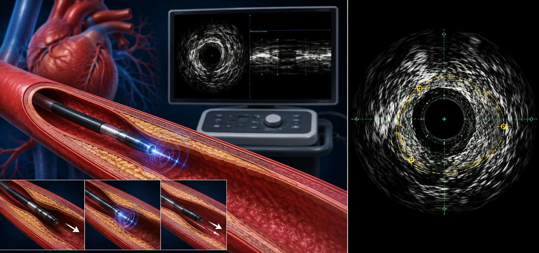
IVUS and OCT Guided Coronary Intervention
IVUS (intravascular ultrasound) and OCT (optical coherence tomography) are imaging techniques used during certain coronary procedures to assess plaque, vessel size and stent placement in greater detail than angiography alone. At Dr Sandeep's Heart Care Center & Labs in Basaveshwaranagar, this imaging may support complex coronary intervention for patients from Vijayanagar and nearby areas.
IVUS / OCT combines intravascular ultrasound and optical coherence imaging concepts used to study the artery from inside and improve planning and optimisation of coronary intervention.
What IVUS / OCT Evaluates
-
The imaging reveals vessel diameter, plaque burden, calcium distribution, and stent expansion.
-
It helps determine where to treat, how large a stent should be, and whether plaque preparation is required.
-
Post-procedure imaging can identify under-expansion, malapposition, or edge problems.
-
These tools improve procedural precision in difficult coronary artery disease.
When IVUS / OCT Is Recommended
-
Left main disease, calcified lesions, long lesions, and bifurcation interventions.
-
Cases where angiography leaves doubt about plaque severity or stent outcome.
-
Patients needing image-guided PCI for better treatment planning.
-
Situations where correcting a previous stent-related issue needs detailed vessel imaging.
Who May Benefit From IVUS / OCT
-
Patients with complex coronary anatomy needing high-precision intervention.
-
Individuals in whom accurate vessel sizing is important for durable stent results.
-
People whose prior procedure needs optimisation or problem-solving.
-
Patients who may benefit from imaging-guided improvement in PCI quality.
Benefits of IVUS / OCT
-
Provides clearer diagnosis and more confident treatment decisions.
-
Helps identify heart problems earlier, before symptoms become serious.
-
Reduces uncertainty when chest pain, breathlessness, or palpitations need explanation.
-
Guides follow-up care, medication changes, and preventive heart planning.
Book a IVUS / OCT Evaluation
Consult Dr Sandeep's Heart Care Center & Labs to know whether IVUS / OCT is the right next step for your symptoms or ongoing heart condition. Timely testing helps clarify diagnosis, guide treatment, and support safer follow-up care.
Dr Sandeep's Heart Care Center & Labs is located in Basaveshwaranagar and is easily accessible for patients travelling from Rajajinagar, Vijayanagar, Magadi Road,Malleshwaram, Mahalakshmipuram, Mahakashmi Layout, Peenya, Yeshwanthpur, Nandini Layout, Dasarahalli and surrounding areas who are looking for a cardiologist for ongoing heart disease management, BP control, or preventive cardiac care.
Frequently Asked Questions
What is the main difference between IVUS and OCT?
IVUS uses ultrasound waves and can assess larger, more calcified vessels well, while OCT uses light for higher-resolution images, particularly useful for detailed plaque and stent-edge evaluation. The choice depends on the specific clinical situation.
Will my cardiologist decide which one to use, or can I choose?
Your interventional cardiologist selects the imaging technique — IVUS, OCT, or neither — based on your coronary anatomy and the specific questions that need to be answered during your procedure.
Does IVUS/OCT guarantee a perfect stent result?
No. While IVUS/OCT can improve the accuracy of stent sizing and placement, no imaging technique can guarantee a specific outcome, as this also depends on individual anatomy and overall health.
Is IVUS/OCT used only during angioplasty, or also afterward?
It can be used both before stenting, to plan treatment, and after stenting, to confirm the stent is well-positioned and fully expanded, depending on the case.
How is IVUS/OCT different from a coronary angiogram?
A coronary angiogram gives an outside view of the artery using dye and X-ray. IVUS and OCT give a detailed inside view of the vessel wall itself, offering additional information the angiogram alone cannot show.
Who typically needs IVUS or OCT guidance during a procedure?
Patients with complex coronary anatomy — such as left main disease, long lesions, heavy calcification, or bifurcation lesions — are more likely to benefit from IVUS or OCT guidance, based on the treating cardiologist's assessment.
.png)